About the Department
Magnetic resonance imaging is a research method that allows you to get a detailed picture of the state of human organs without internal intervention. Since the principle of the device is based on magnetic fields, the research process is absolutely safe from the point of view of ionizing radiation due to its absence.
Nowadays, magnetic resonance imaging is one of the most informative methods of research and diagnosis of diseases of the central nervous system, osteomuscular, articular and other human systems.
Services of the Department
- Magnetic resonance tractography of the brain is a diagnostic method based on diffusion-tensor magnetic resonance imaging, which allows visualizing the orientation and integrity of the brain pathways in vivo.
- Tractography is used in the diagnosis of axonal injuries in chronic brain ischemia, moto neuron diseases, multiple sclerosis, acute disseminated encephalomyelitis, brain tumors and CNS development abnormalities, cortical infarcts.
- Magnetic resonance neuroimaging in epilepsy and convulsive syndrome. It allows to identify a pathological focus (the so-called “epi-focus”) in the brain structure, to determine the tactics of patient management and selection of a treatment method (surgically or conservatively).
- 1H Magnetic resonance-spectroscopy of the brain. Proton spectroscopy allows noninvasively in vivo to obtain information about the chemical composition of the brain substance – the main metabolites. This method allows you to detect pathological changes in the structures of the brain in the early stages of the disease.
- Multimetric program PI-RADS v2 on magnetic resonance imaging (MRI) with a magnetic field strength of 3.0 T.
- Prostate cancer is detected in 4-7% of cases among men aged 50 years and older, who do not have urological symptoms and genitourinary system diseases history. This technique is implemented for the first time in Kazakhstan at the Presidential Hospital and to this day is the only one on the territory of our country, the use of a program with dynamic contrast study with high precision allows detecting pathological lesions in the prostate gland malignancy with accuracy up to 90-95%.
- Magnetic resonance enterography is an innovative method for diagnosing diseases of the small intestine, which currently does not have any competitive research methods that provide information about the state of the small intestine and the surrounding fiber in a non-invasive way, without painful examinations and multiple tests.
Doctors of the Department
 |
Elmira Yelshinbayeva Chief of the Department PhD The Highest category Work experience: 19 years |
 |
Igor Berestyuk Doctor of CT & MRI The Highest category Work experience: 18 years |
 |
Aleksandr Dun Doctor of CT & MRI The First category Work experience: 17 years |
 |
Rayma Kapi Doctor of CT & MRI Master of Medicine The Highest category Work experience: 24 years |
 |
Damilya Mardenkyzy Doctor of CT & MRI PhD The First category Work experience: 13 years |
 |
Aygerim Suygenbayeva Doctor of CT & MRI The First category Work experience: 13 years |
 |
Saltanat Gadylkan Doctor of CT & MRI Work experience: 4 years |
 |
Manar Khoishybai Doctor of CT & MRI Work experience: 3 years |
 |
Takhir Tashpulatov Doctor of CT & MRI Work experience: 4 years |
 |
Shyngys Ibrayev |
 |
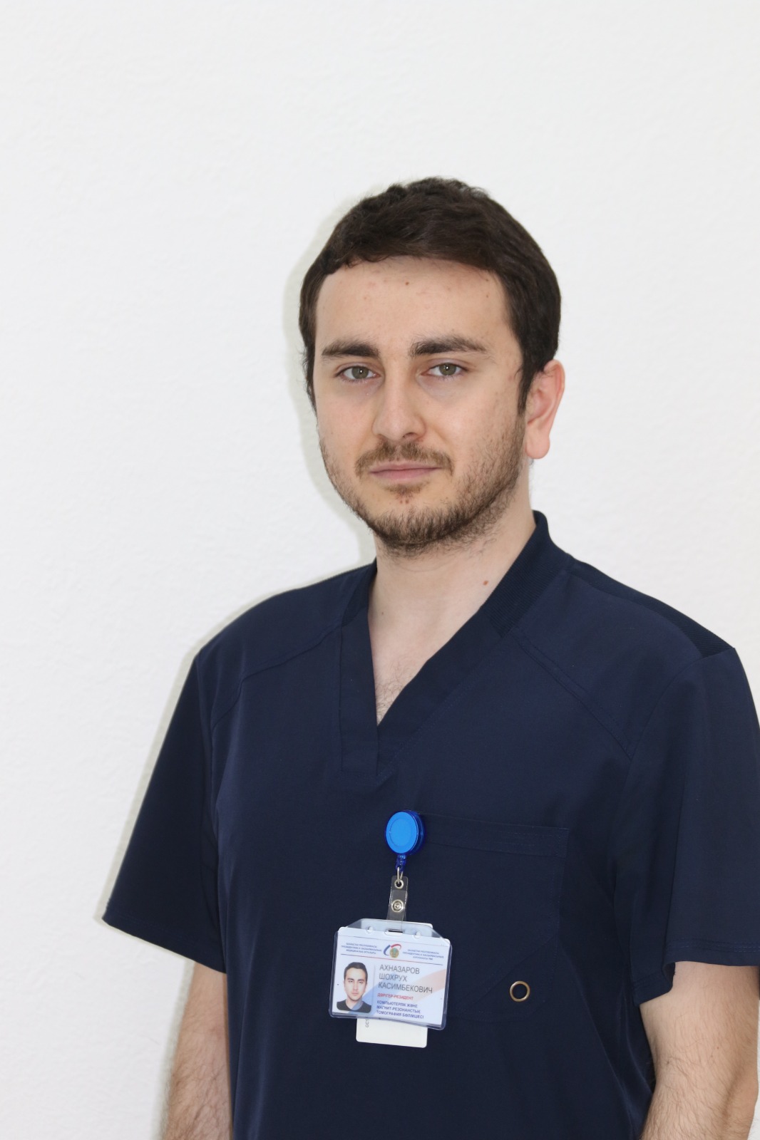
Shokhrukh Ahnazarov |
 |
Damir Bekkozhin |
Contacts of the Department
8 (7172) 70 78 88
8 (7172) 70 79 36
8 (7172) 70-79-34
+ 7 701 534 95 98
Working hours
Mon-Sat: 8:00am. – 08:00pm.


